Anatomy is one of the first and most important subjects in healthcare education. Before students can understand disease, diagnosis, treatment, movement, imaging, or patient care, they need a clear understanding of how the human body is structured. This is not always easy to achieve through textbooks and classroom lectures alone. Traditional anatomy learning has always depended on diagrams, charts, cadaver labs, physical models, and instructor-led explanations. These methods are valuable, but they can sometimes make anatomy feel difficult to visualize. The human body is three-dimensional, and students need to understand how organs, muscles, bones, nerves, and vessels are placed in relation to one another. This is where a virtual anatomy model becomes highly useful. It gives healthcare students a more visual and interactive way to study the body.
What Is a Virtual Anatomy Model?
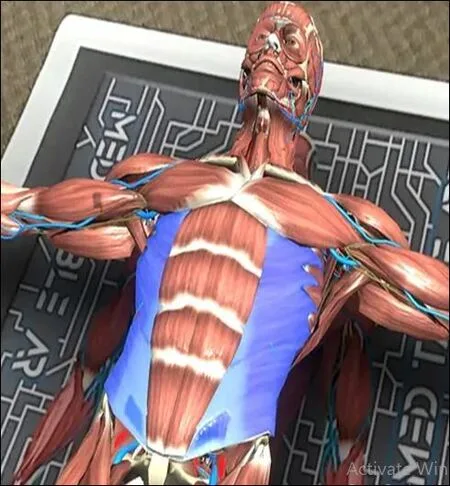
A virtual anatomy model is a digital 3D representation of the human body. It allows students and educators to view anatomical structures in a more detailed and interactive format than flat textbook images.
With this type of model, students can study body systems, organs, bones, muscles, and other structures from multiple angles. They can zoom in, rotate the model, isolate specific parts, and better understand how everything fits together.
This approach is especially useful because anatomy is not just about memorizing names. Students also need to understand position, function, depth, movement, and relationships between structures. A digital model makes these details easier to see and understand.
Why Healthcare Education Needs Better Anatomy Tools
Healthcare students often find anatomy challenging because it involves a large amount of information and complex spatial understanding. A single diagram may show one view of an organ or system, but it may not clearly show how that structure looks from the side, behind, above, or within the body.
For example, understanding the path of a nerve, the placement of internal organs, or the connection between muscles and bones becomes much easier when students can view the structure in 3D. This helps them move beyond basic memorization and develop a more practical understanding of the human body.
Digital anatomy tools support this process by making learning more visual, repeatable, and easier to connect with real healthcare situations. They do not replace textbooks, cadaver labs, or instructor guidance. Instead, they strengthen these methods by giving students another way to understand difficult concepts.
Making Anatomy Easier to Visualize
One of the biggest advantages of using a virtual anatomy model is that it helps students understand the body as a three-dimensional system. This is important because healthcare professionals must think in 3D when reading scans, examining patients, understanding injuries, or learning procedures.
Textbook diagrams are useful for basic learning, but they often simplify structures. In real life, body parts overlap, curve, connect, and sit at different depths. A 3D model allows students to explore these relationships more clearly.
This is especially helpful for subjects such as the cardiovascular system, nervous system, musculoskeletal system, and internal organs. Students can see where structures are located, how they connect, and how they relate to nearby body parts. This makes the learning experience more practical and easier to retain.
Supporting More Interactive Learning
Anatomy learning becomes more effective when students are actively involved. When learners can interact with a model, they are more likely to stay engaged and understand the concept being taught.
Interactive anatomy tools allow students to explore instead of only observe. They can move around a structure, look closer at a specific area, compare systems, and repeat the process until the concept becomes clear. This kind of learning is especially helpful for students who struggle to understand anatomy through reading alone.
MedTableAR supports this style of learning by using augmented reality and 3D anatomical visualization. Its platform is designed to help students explore body systems in a more hands-on way, making anatomy lessons more engaging and easier to follow.
Conclusion
A strong understanding of anatomy is essential for every healthcare student. However, anatomy can be difficult to learn when students rely only on flat diagrams, lectures, or limited lab access. The subject becomes easier to understand when learners can see the body in a more visual, interactive, and three-dimensional way. A virtual anatomy model helps bridge this gap. It supports clearer visualization, better engagement, repeated practice, and stronger classroom teaching. It also works well alongside traditional anatomy resources, making it a practical addition rather than a replacement.
Why a Virtual Anatomy Model Is Essential for Healthcare Education
Anatomy is one of the first and most important subjects in healthcare education. Before students can understand disease, diagnosis, treatment, movement, imaging, or patient care, they need a clear understanding of how the human body is structured. This is not always easy to achieve through textbooks
MedTable AR
·
3 min read
·
85 Buzz