
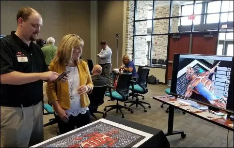
A successful anatomy classroom needs more than detailed content. It needs tools that students can explore together without complicated setup. An interactive 3D anatomy station combines augmented reality, portable devices, a printed body map, and shared display options. This coordinated approach can turn lectures into investigations while helping educators explain structures, body planes, and physiological processes more clearly.
Start With a Classroom-Ready Setup
Introducing educational technology can be difficult when teachers must purchase separate devices, install applications, and solve compatibility problems. A coordinated package reduces these barriers.
A practical AR anatomy setup may include:
● Pre-programmed tablets
● A durable mat with a printed body map
● A protective roll-up case
● A classroom software license
● Product warranty coverage
● A screencast device
● Access to major organs and body systems
With the essential components prepared, educators can spend less time managing equipment and more time guiding learning.
Turn a Printed Map Into a Virtual Body
The printed body map gives the augmented reality application a physical reference point. Students can view digital anatomical structures as if they are positioned within the classroom.
They may be able to:
● Rotate the body
● Zoom in on small details
● Isolate an organ
● Remove anatomical layers
● Perform a virtual dissection
● Examine posterior views
● Compare body planes
● Observe real-time processes
These controls make the learning experience active. Students are not limited to the angle or depth chosen by a textbook illustrator.
Organize Students for Shared Exploration
An interactive 3D anatomy station can support several classroom formats. A teacher may lead a whole-class demonstration, assign students to small groups, or create rotating learning stations.
A practical group lesson could follow this sequence:
1. Introduce the body system.
2. Assign each group a structure or process.
3. Let students explore using a tablet.
4. Ask groups to record key observations.
5. Display the model on a smartboard.
6. Invite students to explain their findings.
7. Finish with a short review activity.
This format encourages observation, communication, and peer learning.
Use the Smartboard for Guided Teaching
A screencast device can display the tablet’s content on a smartboard or secondary monitor. This is valuable when the teacher needs every student to follow the same view.
The educator can highlight an organ, rotate the model, remove a layer, or demonstrate blood flow as they explain each stage. Students can answer questions from their seats before exploring the content in smaller groups.
Shared display also helps classes review difficult material without crowding around one device.
Teach Structure and Function Together
Anatomy lessons often separate structure from function. Interactive technology can bring them together.
Students might locate the heart, examine its position, and then observe blood moving through the circulatory system. They can identify digestive organs before following a dynamic process. They may compare muscles and bones before discussing movement.
This connection helps learners understand why structures are shaped and positioned as they are.
Choose Technology Teachers Can Actually Use
Advanced features have limited value if the interface is confusing. Educators should look for intuitive controls, clear instructions, accurate models, and content suitable for their students’ knowledge level.
Schools should also consider:
● Setup and storage requirements
● Device durability
● License length
● Warranty coverage
● Technical assistance
● Curriculum compatibility
● Options for future content
● Ease of cleaning and transport
The purpose is not to add technology for its own sake. It is to create a dependable teaching resource that fits normal classroom routines.
Make Interactive Learning Part of the Curriculum
A coordinated, interactive 3D anatomy setup can easily transition between teacher demonstrations, small-group investigations, and whole-class review. By combining detailed models, virtual dissection, real-time processes, body planes, portable tablets, and a smartboard display, this approach gives educators multiple ways to teach the same concept. For academies seeking a classroom-ready AR system designed for engaging anatomy instruction, MedTableAR offers a practical foundation for immersive learning.