Zygomatic dental implants have revolutionized how clinicians treat patients with severe maxillary bone loss. For individuals who were once told they lacked sufficient bone for traditional implants, zygomatic implants—anchored in the dense zygomatic (cheek) bone—offer a powerful, life-changing alternative. Yet, despite their remarkable success rates, these procedures are among the most complex in implant dentistry. This is where 3D imaging and digital planning play a transformative role, helping ensure precision, safety, and predictable outcomes.
In this article, we’ll explore how advanced imaging and digital tools are redefining the way surgeons approach zygomatic dental implant placement—from pre-surgical diagnosis to final restoration.

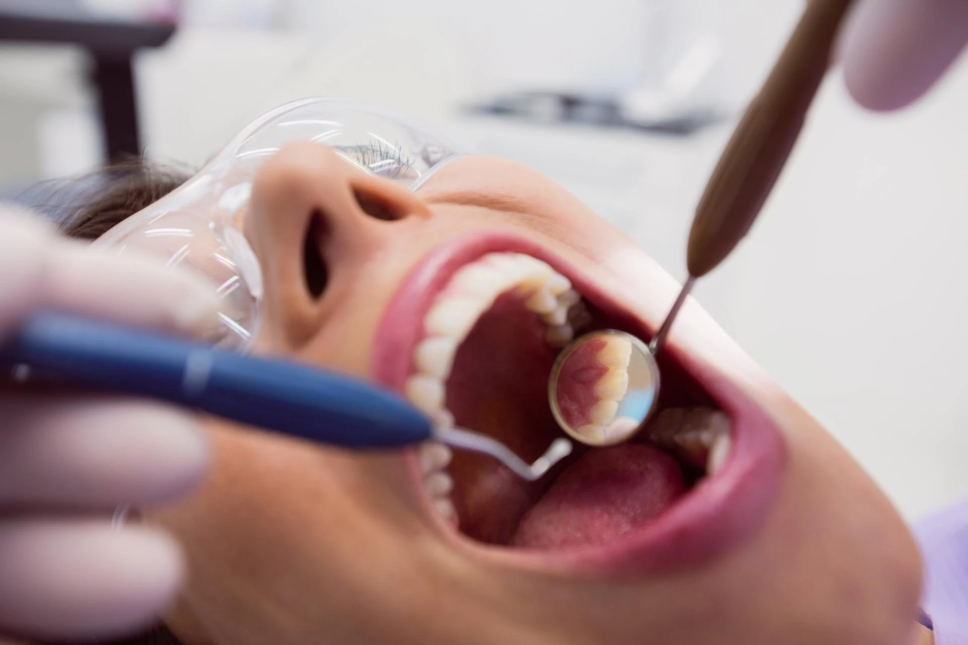
1. The Complexity of Zygomatic Implant Surgery
Unlike conventional dental implants that anchor into the jawbone, zygomatic implants are much longer and traverse the maxillary sinus to engage the zygomatic bone. This anatomical complexity makes traditional two-dimensional X-rays inadequate for planning. Critical structures such as the maxillary sinus, nasal cavity, and orbital floor lie in close proximity to the surgical pathway. Any deviation from the ideal trajectory could result in complications or compromised function.
Therefore, precise visualization of the patient’s anatomy is essential. This is exactly what 3D imaging technologies—such as cone beam computed tomography (CBCT)—offer. With a detailed three-dimensional view, surgeons can plan every aspect of the procedure before ever picking up a drill.
2. How 3D Imaging Enhances Diagnosis and Assessment
3D imaging is now considered a standard of care in implantology, but it’s especially indispensable for zygomatic cases. Using CBCT scans, clinicians can:
- Assess bone quantity and quality: CBCT imaging provides accurate measurements of bone density and volume in both the zygomatic and residual maxillary regions.
- Identify sinus morphology: Every patient’s sinus anatomy is unique. 3D scans reveal variations and potential sinus pathologies that could affect implant trajectory.
- Evaluate adjacent structures: The relationship between the zygomatic bone, orbit, and nasal cavity can be examined to ensure implant safety and avoid nerve or sinus complications.
- Plan for symmetry: When performing full-arch or bilateral procedures, CBCT images allow for comparison between the left and right zygomatic bones to achieve aesthetic and functional harmony.
This level of diagnostic precision not only minimizes surgical risks but also allows clinicians to select the optimal implant length, angulation, and entry point.
3. The Power of Digital Planning Software
Once CBCT data is captured, it can be imported into digital implant planning software such as NobelClinician, coDiagnostiX, or Blue Sky Plan. These programs allow the surgeon to simulate implant placement virtually, testing different angles and trajectories in a 3D environment.
Key advantages of digital planning include:
- Virtual implant placement: Surgeons can pre-determine the exact implant position, depth, and orientation, ensuring a prosthetically driven outcome.
- Integration with intraoral scans: By merging CBCT data with digital impressions, clinicians can visualize both bone and soft tissue structures. This fusion of data supports the design of restorations that fit perfectly with the underlying anatomy.
- Collision detection: The software automatically flags any potential interference with vital structures such as the sinus membrane or orbit.
- Enhanced communication: Digital files can be shared with prosthodontists, lab technicians, and even patients, improving collaboration and understanding of the procedure.
This digital “blueprint” reduces guesswork and ensures that everyone involved in the treatment process works from the same accurate, data-driven plan.
4. Guided Surgery: Turning Plans into Precision
The next evolution after digital planning is computer-guided zygomatic implant surgery. Using the digital plan, surgical guides or navigation systems can help transfer the virtual design into the patient’s mouth with millimeter accuracy.
There are two main approaches:
- Static guided surgery: A physical surgical guide—3D-printed from the digital plan—is placed in the patient’s mouth during surgery. It directs the drilling path and implant insertion angle.
- Dynamic navigation systems: These real-time, GPS-like systems track the drill’s position relative to the patient’s anatomy on a monitor, allowing surgeons to adjust their movements dynamically.
Both techniques have demonstrated improvements in surgical accuracy, reduced operative time, and lower postoperative complications. For zygomatic implants, where even minor deviations can have major consequences, guided technology enhances both safety and predictability.
5. Benefits for Patients and Clinicians
The integration of 3D imaging and digital planning offers numerous benefits for both patients and clinicians:
- Shorter treatment times: Efficient pre-surgical planning allows for faster, more confident surgery and often immediate loading of the prosthesis.
- Less invasive approach: Detailed imaging reduces the need for exploratory surgery and extensive flap elevation, leading to quicker recovery.
- Greater precision and safety: Digital tools minimize the risk of implant misplacement or sinus perforation.
- Improved aesthetic outcomes: The prosthetically driven approach ensures the final restoration aligns perfectly with the patient’s facial structure.
- Enhanced patient confidence: When patients can see their digital treatment plan and understand the process, their trust and acceptance increase significantly.
6. The Future of Digital Zygomatic Implantology
As technology continues to advance, digital workflows are becoming even more seamless. Artificial intelligence (AI) is being integrated into implant planning software to automate landmark identification and suggest optimal implant positioning. Augmented reality (AR) tools are emerging to provide real-time visual overlays during surgery. These innovations will continue to push the boundaries of what’s possible in zygomatic implantology.
Ultimately, the fusion of surgical expertise with cutting-edge digital technology represents the future of complex implant dentistry—delivering safer, faster, and more predictable outcomes for patients who once had no viable solution.
Conclusion
3D imaging and digital planning have redefined the standards of care in zygomatic implant surgery. What was once considered a highly specialized and risky procedure can now be performed with remarkable accuracy and confidence. By combining precise anatomical visualization with advanced digital workflows, clinicians can restore smiles, function, and quality of life to patients who might otherwise remain edentulous.
The result is not just a technical triumph—it’s a powerful example of how digital innovation continues to transform the art and science of implant dentistry.