Learning anatomy is fundamentally a 3D problem. Yet many students still do most of their learning from 2D diagrams, slides, and static models. That mismatch is where augmented reality (AR) becomes genuinely useful. AR places interactive 3D anatomy into the learner’s real space, so students can rotate structures, study relationships between systems, and connect “where it is” to “why it matters” in patient care. For educators, the best AR solutions are the ones that work in real classrooms: fast setup, clear visuals, and group-friendly teaching. MedTableAR is built around that practical need, using tablet-based AR and an anatomy mat to anchor the experience so learners can explore full-body systems and major organs with consistent orientation.
Why AR Improves Anatomy Education In Healthcare Programs
The hardest part of anatomy education usually isn’t memorizing names—it’s understanding spatial relationships and applying them clinically. Students need to grasp depth, layers, planes, and pathways (vessels, nerves, airway, GI flow) under time pressure. AR helps because it turns anatomy from “imagine this in 3D” into “see and manipulate it in 3D.”
With AR, learners can:
● rotate and zoom structures to match the viewpoint they’re being taught (or tested) from,
● isolate systems to reduce visual overload,
● and revisit confusing regions quickly without waiting for the next lab.
This is especially relevant across medical, nursing, and allied health pathways because all three groups rely on spatial accuracy—just in different ways. Medical learners often need deeper structural detail and clinical correlation; nursing learners benefit from clear body-system relationships tied to assessment and procedures; allied health learners often need functional anatomy that supports movement, imaging, emergency response, or rehab.
Stronger 3D Understanding, Faster Clinical Transfer
AR’s biggest advantage is simple: it makes spatial anatomy easier to learn correctly the first time. When students can rotate an organ or trace the route of a vessel interactively, they build a mental map that’s closer to what they’ll need in practice.
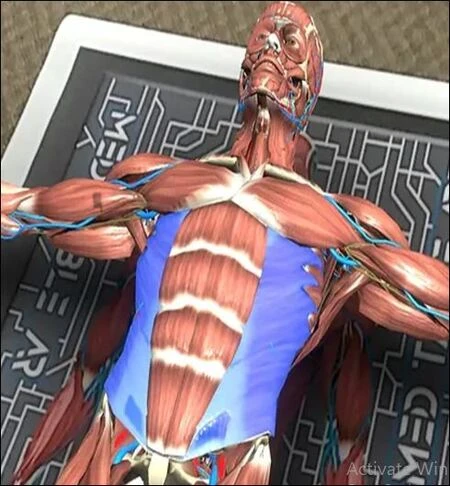
MedTableAR emphasizes interactive 3D exploration of body systems and major organs, helping students examine structures from different angles and in a more realistic spatial context than flat media can provide.
That directly supports common learning outcomes such as:
● identifying structures reliably (not just recognizing a diagram),
● understanding adjacency (“what sits next to what”),
● and applying anatomy to scenarios (pain referral, injury patterns, procedures, and imaging orientation).
Better Layering And System Relationships Without Losing Context
Anatomy doesn’t come neatly separated into chapters. Systems overlap constantly. Students often struggle because they learn a structure in isolation, then can’t place it back into the full-body context. Good AR experiences let learners move between “big picture” and “detail” quickly—view a system, isolate a region, inspect a structure, then return to whole-body orientation. MedTableAR is designed for system-by-system exploration and includes modules that let learners study interrelations and drill into organs with scalable models. This “zoom in, zoom out” flow is one of the most efficient ways to reduce confusion, especially for learners who find anatomy visually overwhelming early on.
Making Structure-Function Links Easier To Understand
Healthcare students don’t learn anatomy as trivia—they learn it to understand function. AR supports that by showing movement and processes, not just shapes. MedTableAR materials highlight motion demonstrations (for example, blood flow through the heart) to help connect structures to physiological behavior.
That matters in:
● nursing education, where anatomy and physiology are tightly linked to assessment and patient monitoring,
● allied health education, where function and mechanism drive decision-making,
● and early medical education, where structure-function understanding is the base for pathology later.
More Effective Group Teaching In Labs And Classrooms
One common failure mode of new teaching tech is that it works for one student on one device—but not for an instructor managing a room. AR becomes far more valuable when it supports group instruction and quick collaboration. MedTableAR is built to accommodate small-group use and classroom display: its FAQ notes the system can support up to six students using the included tablets (typically in pairs per tablet), and it can be shown on a smartboard/monitor using screencasting.
What AR Does Not Replace (And How To Set Expectations)
AR improves clarity and engagement, but it shouldn’t be framed as a replacement for everything. It won’t teach tissue handling or dissection techniques on its own. It also won’t automatically improve outcomes if it’s used without structure (no guided tasks, no checks for understanding). Where AR consistently earns its place is when it reduces avoidable friction in anatomy education—the time students spend confused about orientation, layers, and relationships—and converts that time into meaningful practice.
Conclusion
AR improves anatomy learning because it matches the reality of the subject: three-dimensional structures, layered systems, and clinically meaningful spatial relationships. For medical, nursing, and allied health students, that means faster comprehension, fewer foundational gaps, and better transfer from “I memorized it” to “I can use it.” MedTableAR aligns well with that practical goal by providing interactive 3D models of body systems and organs, support for group learning and classroom display, and reinforcement tools like quizzes and motion demonstrations. In short, AR works best when it is classroom-ready—and when it strengthens anatomy education in ways students can feel immediately: clearer visuals, stronger understanding, and smoother learning from day one.