

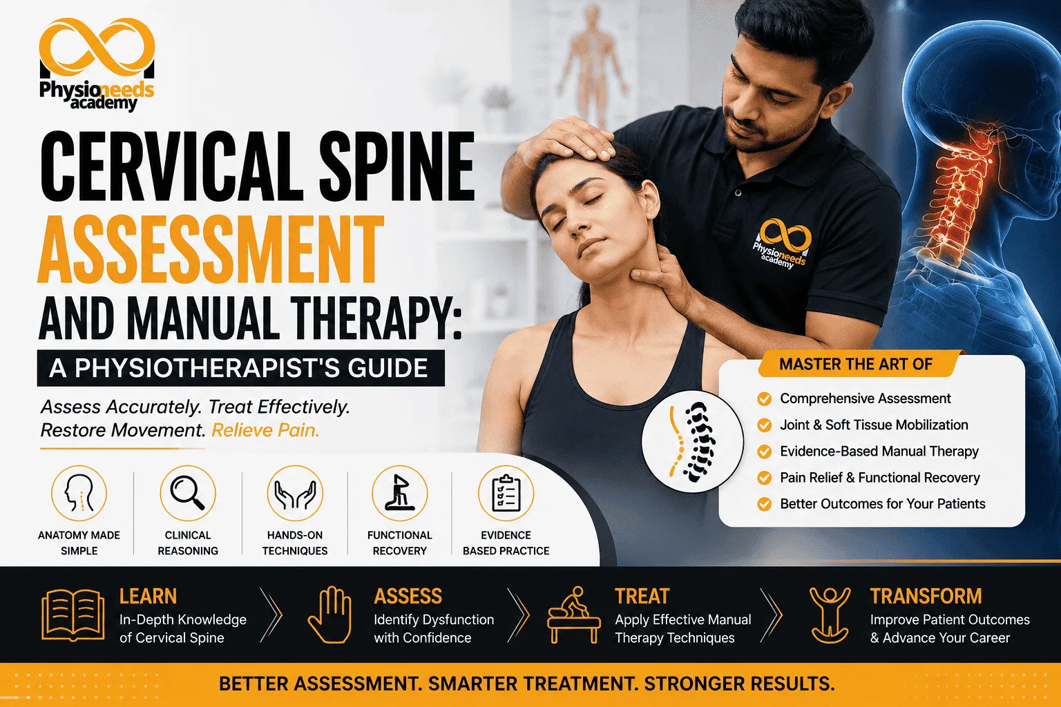
Neck pain affects a significant portion of the population at some point in life, and the cervical spine's complex anatomy makes it one of the easiest regions to assess poorly. After two decades managing cervical spine disorders across orthopaedic and sports medicine settings, I've learned that rushing into treatment without a structured assessment is the biggest mistake physiotherapists make with this region.
This guide covers cervical spine biomechanics, a step-by-step assessment framework, the special tests that genuinely add clinical value, manual therapy techniques, and the safety screening that must precede any hands-on treatment.
How do physiotherapists assess and treat cervical spine dysfunction? Assessment involves a structured subjective history, observation, active and passive range of motion testing, neurological screening, and special tests like the Spurling test and ULNT. Treatment combines evidence-based manual therapy — joint mobilisation, soft tissue work — with targeted exercise therapy, guided throughout by thorough safety screening for red flags.
Understanding Cervical Spine Anatomy and Biomechanics The cervical spine consists of seven vertebrae (C1–C7), divided functionally into the upper cervical spine (C0–C2), responsible for roughly half of cervical rotation, and the lower cervical spine (C3–C7), contributing most of the flexion, extension, and lateral flexion. This division matters clinically, since restricted upper cervical rotation presents very differently from lower cervical segmental stiffness.
Quick Summary: The upper cervical spine (C0–C2) is primarily responsible for rotation, while the lower cervical spine (C3–C7) handles flexion, extension, and lateral flexion. Deep neck flexors and extensors provide dynamic segmental control. Common pain generators include facet joints, discs, muscles, and nerve roots. Coupled movement patterns mean pure single-plane motion is rare.
Muscular support comes from deep stabilisers like longus colli and multifidus, alongside larger superficial muscles such as upper trapezius and levator scapulae, which often become overactive when deep stabilisers underperform. Common pain generators include facet joints, discs, and irritated nerve roots, though presentations frequently overlap, which is why structured assessment matters.
Step-by-Step Clinical Assessment of the Cervical Spine
A thorough cervical assessment builds systematically toward an accurate clinical picture rather than jumping straight to special tests.
Step 1 – Subjective History: onset, mechanism, aggravating/easing factors, red flag screening. Step 2 – Observation and Posture: head position, forward head posture, asymmetry, resting tension. Step 3 – Active Range of Motion (AROM): flexion, extension, rotation, lateral flexion, noting quality and symptom reproduction. Step 4 – Passive Range of Motion (PROM): comparing passive to active findings to identify joint versus muscular limitations. Step 5 – Neurological Examination: myotomes, dermatomes, and reflexes to screen for radicular involvement. Step 6 – Functional Assessment: impact on work, sleep, and daily activities. Step 7 – Clinical Reasoning: synthesising findings into a working diagnosis and treatment plan.
Each step narrows the differential diagnosis progressively, so special tests confirm — rather than replace — reasoning built from earlier steps.
Special Tests Used in Cervical Spine Assessment
The Spurling test is used to identify cervical radiculopathy; a positive result reproduces radicular arm pain, and it carries moderate specificity, so it supports but doesn't confirm a diagnosis on its own. The Upper Limb Neurodynamic Test (ULNT) assesses neural mechanosensitivity by reproducing or worsening arm symptoms, helping differentiate neural pain from musculoskeletal pain. The Cervical Distraction Test checks whether traction relieves symptoms — a reduction in radicular pain supports a radiculopathy diagnosis. The Deep Neck Flexor Endurance Test assesses deep stabiliser endurance, with early fatigue or compensation guiding exercise prescription. VBI Screening screens vascular risk before manual therapy, watching for dizziness, nystagmus, or visual disturbance, and is essential before any upper cervical technique.
No single test should be used in isolation — clustering findings across history, AROM, and multiple special tests produces far more reliable conclusions than relying on any one positive result.
Manual Therapy Techniques for Cervical Dysfunction
Cervical joint mobilisation reduces pain and restores segmental mobility, and is used for mechanical neck pain and facet stiffness. Thoracic mobilisation addresses contributing thoracic stiffness, particularly when reduced cervical rotation is linked to thoracic restriction. Soft tissue mobilisation reduces muscular tension and guarding, especially in the upper trapezius and levator scapulae. Muscle energy techniques improve joint position and mobility where segmental restriction involves muscle guarding. Neural mobilisation reduces nerve mechanosensitivity in patients with radicular symptoms and neural tension signs. Manual traction offers temporary symptom relief and helps assess responsiveness, particularly for radicular pain that responds well to distraction testing.
Technique selection should follow directly from assessment findings rather than habit — a patient with isolated thoracic stiffness limiting cervical rotation, for example, often improves more from thoracic mobilisation than from repeated cervical-focused treatment.
In terms of short-term pain relief, manual therapy often provides faster initial relief, while exercise therapy builds relief more gradually over sessions. For functional improvement, manual therapy offers moderate gains that are often temporary without exercise, whereas exercise therapy delivers strong improvement, particularly for long-term function. Looking at long-term outcomes, manual therapy has limited evidence as a standalone approach, while exercise therapy has better long-term evidence, especially with adherence. On patient adherence, manual therapy is passive and requires ongoing clinic visits, while exercise therapy requires active patient participation. And in terms of evidence strength, manual therapy is a useful adjunct, especially short-term, while exercise therapy has a stronger evidence base for sustained outcomes.
https://www.e-physioneeds.com/courses/A-Z-Spine-6452276ce4b081d895e380e2