Teaching cellular biology often involves asking students to imagine life on a scale that feels abstract or invisible. However, introducing Amoeba proteus into the laboratory transforms these concepts into a captivating, real-time observation of life in flux. Known for its ever-changing shape and unique mode of locomotion, this large protist serves as a primary model for understanding eukaryotic cell structure and behaviour. By providing students with the opportunity to witness a single cell navigate its environment, educators can spark a deep interest in the complex mechanisms that drive even the simplest forms of life.
Observing Pseudopodia and Amoeboid Movement
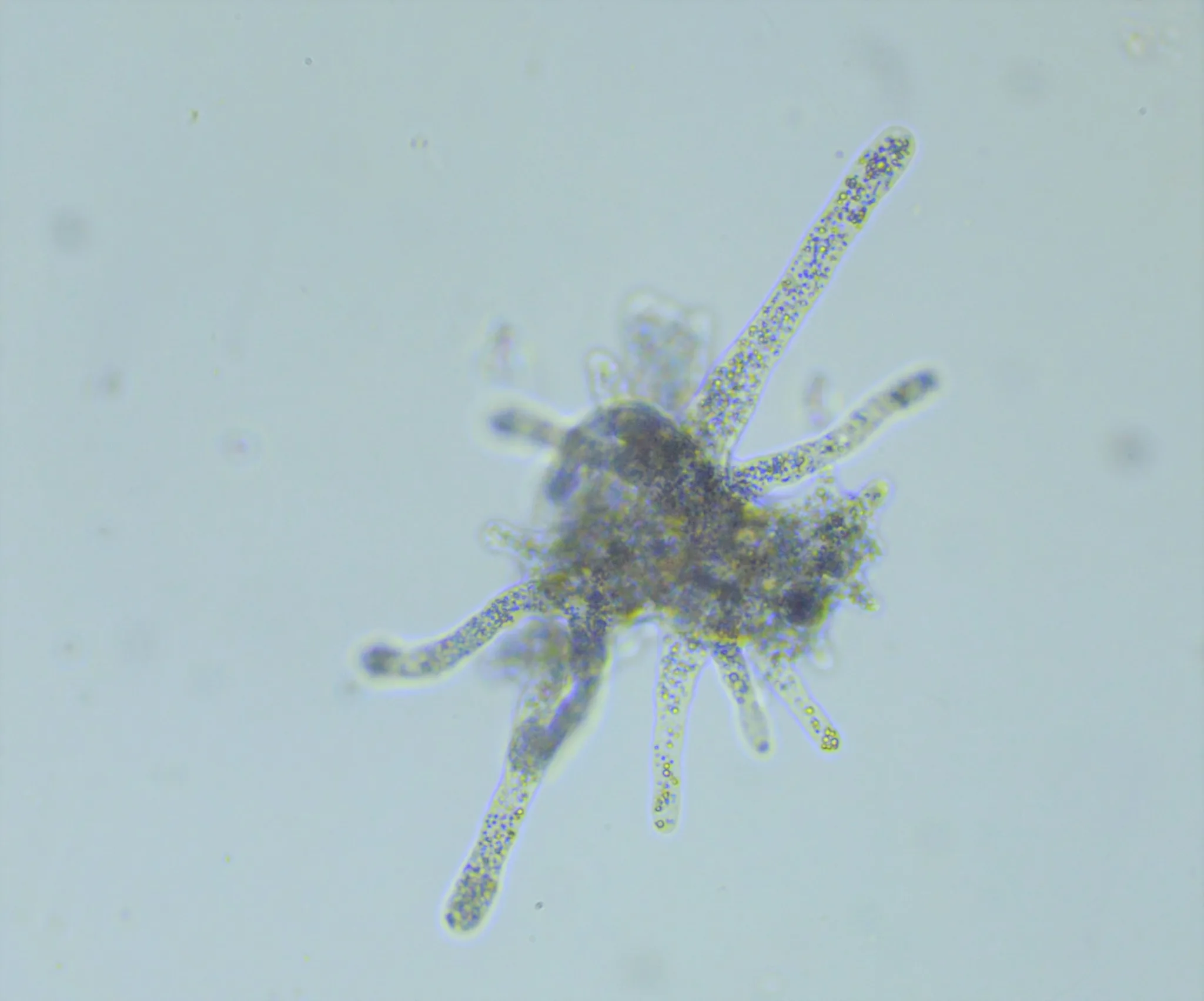
The most distinctive characteristic of Amoeba proteus is its use of pseudopodia, or "false feet," for movement and capturing prey. Under a microscope, students can observe the fluid transition of endoplasm and ectoplasm, a process known as cytoplasmic streaming. This internal flow pushes the cell membrane forward, allowing the amoeba to "crawl" across the slide. Unlike the fixed structures found in prepared microscope slide sets, living specimens demonstrate the dynamic nature of the cytoskeleton in real-time.
To achieve the best results, students should use a cavity slide with a small amount of debris, such as silk fibres or sand grains. This provides the amoeba with a surface to grip and navigate. While a protozoa slide online is excellent for identifying the nucleus and contractile vacuole in high contrast, the living culture is essential for understanding the energetic cost and physical mechanics of locomotion.
Demonstrating Phagocytosis and Nutrient Uptake
In addition to movement, Amoeba proteus offers a spectacular demonstration of phagocytosis. When the amoeba encounters a food source, such as small ciliates or flagellates, it extends its pseudopodia to surround and engulf the prey, forming a food vacuole. This process provides a clear, visible example of how cells interact with their environment to obtain energy. It is a foundational lesson in biology that bridges the gap between basic cell structure and complex metabolic functions.
For a comparative study of microscopic life, educators often introduce other organisms into the lab. For instance, comparing the slow, deliberate movements of the amoeba to the rapid swimming of organisms found in a freshwater life set highlights the diversity of survival strategies in a single drop of water.
Integrating Microscopic and Macroscopic Studies
While the focus remains on the living cell, a comprehensive biology curriculum benefits from integrating various types of specimens. While students watch a living amoeba proteus hunt, they can simultaneously reference an amoeba proteus slide to identify organelles that may be obscured by the movement of the cytoplasm in a live sample. This dual approach—using both living and preserved materials ensures that students develop a high level of competency in microscopy and biological identification.
Furthermore, the study of single-celled organisms often leads to questions about more complex life forms. A lab session might begin with the microscopic world and transition into vertebrate anatomy. For example, a teacher might use frozen specimens for a heart or kidney dissection to show how trillions of specialised cells work together in a way that parallels the functions performed by a single amoeba. Supplementing these lessons with specialised tools, such as an owl pellet dissection mat for ecology units or microscope slide sets for histology, creates a well-rounded and varied learning experience that prevents student fatigue and repetitive lesson structures.
Maintaining Culture Health and Lab Standards
Success in observing protists depends heavily on the quality of the culture and the equipment used. Amoeba proteus is sensitive to temperature changes and chemical contaminants, so using clean, spring water and providing a steady food source of Chilomonas or Colpidium is vital. This level of care introduces students to the responsibilities of laboratory management and the importance of maintaining living systems.
When sourcing materials, working with a specialised biology lab supply ensures that the specimens arrive in peak condition, with high cell counts and minimal contamination. This reliability is crucial for timed classroom sessions where every minute counts. Whether the lesson involves the slow crawl of a protist or the detailed analysis of fungi microscope slides, having professional-grade materials is the difference between a failed experiment and a breakthrough moment of student discovery.
Blades Biological: Precision Science Education
Blades Biological supports educators with reliable, high-quality materials designed for effective teaching. By choosing living amoeba proteus cultures, schools provide students with vibrant specimens that facilitate dynamic learning experiences. Supplementary resources, such as science education specimens and biological equipment for sale, equip classrooms for hands-on exploration of cellular behaviour, nutrient uptake, and microscopic ecology.
Blades Biological ensures safe, accurate, and reproducible materials. From live protists to prepared microscope slide sets and fungi microscope slides, the company enables educators to integrate interactive biology lessons seamlessly into the curriculum. These tools foster curiosity, reinforce theoretical knowledge, and encourage students to develop analytical and observational skills.
Bring the microscopic world to life in your classroom, explore Blades Biological’s range of living protist cultures and educational specimens today.