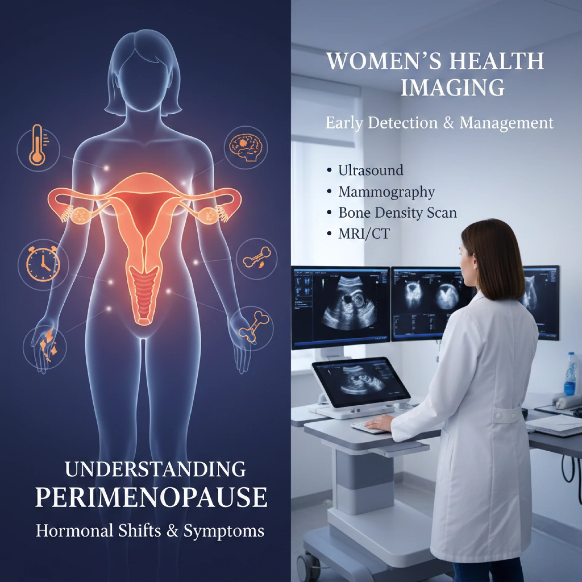
Perimenopause is a natural phase of life that many women experience, typically beginning in their 40s but sometimes earlier. It marks the transitional period before menopause, when the ovaries gradually produce less estrogen and menstrual cycles become irregular. While perimenopause is a normal part of aging, the changes it brings can be confusing, uncomfortable, and at times concerning. Understanding what’s happening in your body and knowing when to seek medical support can make this transition much more manageable.
One powerful tool in modern gynecologic care is women’s health imaging. This includes diagnostic tests like ultrasounds, mammograms, and bone density scans that help doctors evaluate reproductive organs, detect abnormalities, and monitor conditions that may arise during perimenopause and beyond. Advances in women’s health imaging have transformed how clinicians diagnose and treat conditions that were once difficult to detect until symptoms became severe.
In this article, we’ll explore what perimenopause is, the common symptoms associated with it, why women’s health imaging plays a critical role in diagnosis and care, and how women can empower themselves to navigate this stage with confidence.
What Is Perimenopause?
Perimenopause is the transitional period leading up to menopause, defined as the point when a woman has gone 12 consecutive months without a menstrual period. This transition can last anywhere from a few months to several years, and it is characterized by fluctuating hormone levels especially estrogen and progesterone.
During perimenopause, the body may respond to hormonal changes in a variety of ways. Some women experience mild symptoms, while others face significant disruptions in their day-to-day life.
Common symptoms include:
- Irregular menstrual cycles: Periods may become longer, shorter, heavier, or lighter, and sometimes skip months altogether.
- Hot flashes and night sweats: Sudden feelings of warmth or intense sweating, particularly at night.
- Mood changes: Increased irritability, anxiety, or depression.
- Sleep disturbances: Difficulty falling asleep or staying asleep.
- Vaginal dryness: Changes in vaginal tissue due to decreased estrogen.
- Decreased libido: Reduced sexual desire.
- Cognitive changes: Challenges with concentration or memory.
Because perimenopause overlaps with age ranges where other health concerns (such as fibroids, endometriosis, or ovarian cysts) may also emerge, differentiating between normal transitional symptoms and other conditions becomes essential.
Women’s Health Imaging: A Vital Resource in Perimenopause Care
Women’s health imaging refers to a suite of diagnostic tools that allow clinicians to look inside the body and assess the health of reproductive organs, breasts, bones, and surrounding tissues. These imaging techniques help doctors gather accurate information to diagnose, monitor, and treat gynecologic conditions many of which can be especially relevant during perimenopause.
Let’s walk through some of the key imaging tools used in women’s health care:
Ultrasound Imaging
Ultrasound uses sound waves to create images of internal organs without radiation. It is often the first diagnostic step when evaluating symptoms such as pelvic pain, abnormal bleeding, or suspected ovarian cysts.
During perimenopause, ultrasounds can help:
- Detect fibroids or polyps
- Evaluate ovarian cysts
- Monitor endometrial thickness
- Assess pelvic organs for structural abnormalities
This imaging tool is quick, safe, and effective, making it a cornerstone of women’s health imaging.
Mammograms
Mammography is a specialized X-ray exam used to screen for breast cancer. As women reach their 40s and beyond a time that often overlaps with perimenopause routine mammograms become increasingly important.
Mammograms can detect:
- Early signs of breast cancer before symptoms appear
- Calcifications or suspicious masses
- Changes in breast tissue density
Regular mammography is a key component of preventive women’s health care, especially during midlife transitions.
Bone Density Scans
During perimenopause, the decline in estrogen can contribute to bone density loss, increasing the risk of osteoporosis. A bone density scan (DEXA) measures bone strength and helps doctors assess fracture risk.
This form of women’s health imaging is particularly crucial because it:
- Detects early bone loss before fractures occur
- Guides treatment decisions to preserve bone health
- Monitors the effectiveness of osteoporosis therapies
By incorporating bone density testing into routine care, women can proactively manage long-term skeletal health.
When Is Women’s Health Imaging Recommended?
Not all women will need advanced imaging during perimenopause, but there are specific situations when it becomes especially valuable. Your doctor may recommend imaging if you experience:
- Unusual vaginal bleeding: Heavy or irregular bleeding that deviates from your normal pattern
- Persistent pelvic pain: Discomfort that doesn’t improve with time
- Abnormal mammogram results: Follow-up imaging to investigate breast abnormalities
- Family history of cancer: Increased surveillance based on risk factors
- Signs of bone loss: Height loss or fractures that raise concern for osteoporosis
Imaging helps confirm or rule out conditions that may mimic perimenopausal symptoms, ensuring that you receive the care you need without unnecessary delay.
Empowering Women Through Education and Prevention
While perimenopause is a natural biological process, many women feel unprepared for its physical and emotional effects. Education and proactive health management are powerful tools for navigating this transition with confidence.
Here are a few recommendations:
Track Your Symptoms
Keeping a journal of your symptoms, menstrual cycle changes, sleep patterns, and emotional well-being can help your healthcare provider recognize trends and make informed decisions about imaging or interventions.
Stay Up to Date With Screenings
Routine women’s health imaging, such as mammograms and bone density scans, provides essential insights into your health. Discuss with your provider when these screenings should begin based on your age, family history, and personal risk factors.
Frequently Asked Questions (FAQ)
1. What is the difference between perimenopause and menopause?
Perimenopause is the transitional period leading up to menopause, when hormone levels fluctuate and menstrual cycles become irregular. Menopause is confirmed when a woman has gone 12 consecutive months without a period. Symptoms of perimenopause can begin several years before menopause.
2. How does women’s health imaging help during perimenopause?
Women’s health imaging provides visual insights into organs and tissues that may be causing symptoms. For example, ultrasounds can assess pelvic pain or abnormal bleeding, mammograms can detect early breast changes, and bone density scans can measure bone strength. These tools help clinicians deliver accurate diagnoses and personalized care.
3. At what age should I start mammograms?
Most guidelines recommend starting routine mammograms at age 40, but your doctor may suggest earlier screening based on factors like family history or personal risk. Women’s health imaging is tailored to each individual’s needs, so it’s important to discuss your risk factors with your provider.
4. Can imaging detect conditions besides cancer?
Yes. Women’s health imaging can detect a variety of conditions, including fibroids, ovarian cysts, polyps, bone loss, hormonal changes affecting reproductive organs, and more. Imaging is not only for cancer detection it’s a versatile diagnostic tool.
5. Are ultrasounds safe?
Yes. Ultrasound imaging uses sound waves rather than radiation, making it a safe and widely used method for examining internal organs, including reproductive structures.
6. Does every woman in perimenopause need imaging?
Not necessarily. Imaging is recommended based on individual symptoms, health history, and screening needs. A healthcare provider can help determine which tests are appropriate for you.
7. Can bone density scans prevent osteoporosis?
While bone density scans don’t prevent osteoporosis themselves, they can detect early bone loss so that interventions can begin before fractures occur. Lifestyle changes and medications may be recommended based on your results.
8. How often should I get screened with women’s health imaging?
Screening schedules vary by the type of imaging and personal risk factors. Mammograms are typically recommended annually or biennially beginning at age 40, while bone density scans are guided by age, hormone status, and clinical indicators. Follow your provider’s recommendations for the best schedule.
Perimenopause doesn’t have to be a mystery. By understanding what’s happening in your body and using tools like women’s health imaging when appropriate, you can navigate this chapter of life with confidence, clarity, and peace of mind.