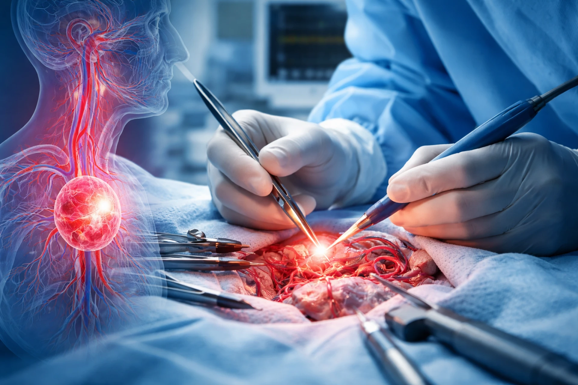
Vascular tumours are abnormal growths that arise from blood vessels or lymphatic vessels. When these tumours occur in complex anatomical regions like the head and neck, accurate diagnosis becomes crucial for effective management. Imaging tests play a vital role in detecting, characterizing, and planning treatment for these tumours. Understanding how different imaging techniques work can help patients and caregivers better navigate the diagnostic process and explore appropriate head and neck vascular tumor treatment options.
What Are Vascular Tumours?
Vascular tumours can be benign, intermediate, or malignant. Common examples include hemangiomas, vascular malformations, and angiosarcomas. In the head and neck region, these tumours may affect areas such as the oral cavity, nasal passages, throat, or salivary glands. Because of the rich blood supply and complex structures in this region, early and precise diagnosis is essential.
Imaging tests help determine:
- The size and extent of the tumour
- Its exact location
- Involvement of nearby tissues or organs
- Blood supply and vascular characteristics
These details are critical when selecting appropriate head and neck vascular tumor treatment options.
Importance of Imaging in Diagnosis
Clinical examination alone is often not sufficient to fully evaluate vascular tumours. Imaging provides a non-invasive way to look inside the body and understand the behaviour of the tumour. It helps in:
- Differentiating between benign and malignant lesions
- Planning surgical or non-surgical treatment
- Monitoring response to therapy
- Detecting recurrence
Each imaging modality has its own strengths, and doctors often use a combination of tests for a comprehensive evaluation.
Common Imaging Tests Used
1. Ultrasound (USG)
Ultrasound is usually the first imaging test recommended, especially for superficial vascular tumours.
How it works:
It uses high-frequency sound waves to create real-time images of internal structures.
Advantages:
- Non-invasive and painless
- No radiation exposure
- Cost-effective
- Useful for evaluating blood flow using Doppler imaging
Role in vascular tumours:
Ultrasound helps identify whether a lesion is solid or cystic and assesses blood flow patterns, which are important indicators of vascular nature.
2. Computed Tomography (CT Scan)
A CT scan provides detailed cross-sectional images of the body using X-rays.
How it works:
Multiple X-ray images are combined to create a 3D view of the tumour and surrounding structures.
Advantages:
- Fast and widely available
- Good for evaluating bone involvement
- Helpful in emergency situations
Role in vascular tumours:
CT scans are particularly useful in assessing tumours located in deeper regions of the head and neck. Contrast-enhanced CT can highlight blood vessels and tumour vascularity, aiding in diagnosis and treatment planning.
3. Magnetic Resonance Imaging (MRI)
MRI is one of the most important imaging tools for vascular tumour detection in the head and neck.
How it works:
It uses strong magnetic fields and radio waves to generate detailed images of soft tissues.
Advantages:
- Excellent soft tissue contrast
- No radiation exposure
- Superior for evaluating tumour extent
Role in vascular tumours:
MRI is highly effective in distinguishing different types of tissues and identifying tumour boundaries. It is especially useful for assessing involvement of muscles, nerves, and blood vessels. MR angiography (MRA) can further visualize blood flow within the tumour.
4. Angiography
Angiography is a specialized imaging technique used to visualize blood vessels.
How it works:
A contrast dye is injected into the blood vessels, and X-ray images are taken to track blood flow.
Advantages:
- Provides detailed visualization of vascular anatomy
- Helps identify feeding arteries and draining veins
Role in vascular tumours:
Angiography is particularly useful for highly vascular tumours. It helps in planning procedures such as embolization, where blood supply to the tumour is reduced before surgery.
5. Positron Emission Tomography (PET Scan)
PET scans are often combined with CT (PET-CT) for better accuracy.
How it works:
A small amount of radioactive tracer is injected into the body, which accumulates in active cells like cancer cells.
Advantages:
- Detects metabolic activity
- Helps identify malignant tumours
- Useful in staging and detecting metastasis
Role in vascular tumours:
PET scans are especially valuable when there is suspicion of cancer. They help determine whether the tumour has spread to other parts of the body.
6. Digital Subtraction Angiography (DSA)
DSA is an advanced form of angiography.
How it works:
It removes background structures from images, leaving a clear view of blood vessels.
Advantages:
- Highly precise vascular imaging
- Useful for interventional procedures
Role in vascular tumours:
DSA is often used before surgical or interventional procedures to map out the tumour’s blood supply in detail.
Choosing the Right Imaging Test
The choice of imaging test depends on several factors:
- Location of the tumour
- Size and suspected type
- Patient’s medical history
- Need for detailed vascular assessment
In many cases, doctors use a combination of ultrasound, MRI, and CT scans to get a complete picture. Advanced imaging like PET scans or angiography is used when more detailed evaluation is required.
Role of Imaging in Treatment Planning
Accurate imaging is essential for selecting the most appropriate head and neck vascular tumor treatment options. These may include:
- Observation for small, benign tumours
- Medication or targeted therapy
- Minimally invasive procedures like embolization
- Surgical removal
- Radiation therapy in certain cases
Imaging helps determine whether surgery is feasible, how extensive it should be, and how to minimize risks such as excessive bleeding.
Monitoring and Follow-Up
Imaging does not stop at diagnosis. It is also used during and after treatment to:
- Monitor tumour response
- Detect any recurrence
- Evaluate complications
Regular follow-up imaging ensures that any changes are detected early, allowing timely intervention.
Conclusion
Imaging tests are a cornerstone in the detection and management of vascular tumours, especially in the complex head and neck region. From initial diagnosis to treatment planning and follow-up, these technologies provide critical insights that guide clinical decisions.
Understanding the role of different imaging modalities can help patients make informed decisions and feel more confident about their care journey. With advancements in medical imaging, doctors can now diagnose vascular tumours more accurately and tailor head and neck vascular tumor treatment options to each individual’s needs.
For those seeking specialized care, consulting an experienced professional—such as the best head and neck cancer surgeon in Ahmedabad—can ensure that imaging findings are interpreted correctly and translated into an effective treatment plan.