In medical science, visualizing the human body in its true complexity has always been a challenge. Textbooks and 2D scans offer limited perspectives, while cadavers—though invaluable—can’t always represent patient-specific anatomy. That’s where the science of 3D surgical models comes in.
Far from being just plastic replicas, these 3D printed anatomical models are powerful tools rooted in imaging data, cutting-edge printing technology, and biomedical precision. Let’s break down the real science behind them—and why surgeons, students, and specialists are turning to companies like Curewith3D for these life-like, hyper-accurate models.
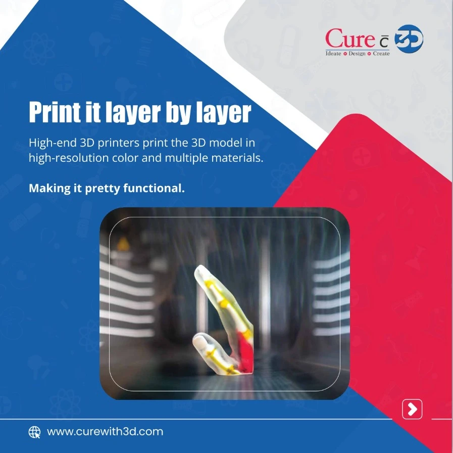
From Scan to Model: The Process
It all starts with medical imaging. CT scans, MRIs, and angiograms generate the raw data. That data is then converted into a 3D file using specialized software, which allows technicians to digitally isolate bones, tissues, or organs. Once the virtual model is perfected, it’s sent to a high-precision 3D printer.
Using biocompatible materials and industry-grade printers, 3D surgical models come to life—layer by intricate layer. The result? A tangible, patient-specific model that reflects every curve, vessel, and variation in anatomy.
Not Just for Display: These Models Save Lives
Unlike standard plastic demos, 3D anatomical models are designed for real-world applications:
- Surgical planning
- Medical education
- Doctor-patient communication
- Implant pre-fitting and trial surgeries
Let’s look at how various anatomical models are making an impact.
Brain Anatomy Model: Navigating Complexity
The brain’s structure is one of the most complex in the human body. A brain anatomy model helps neurosurgeons plan delicate surgeries by providing a full 3D view of tumors, aneurysms, or vascular anomalies. For patients, it simplifies understanding of conditions that often sound intimidating in 2D scans.
Anatomical Heart Model: For Cardiologists and Beyond
A patient-specific anatomical heart model can replicate congenital defects, arterial blockages, or valve irregularities. Cardiologists can use these to explain procedures like stenting or bypass surgery, while surgical teams practice complex repairs before entering the operating room.
Eye and Ear Anatomy Models: Tiny Structures, Big Impact
With ultra-detailed eye anatomy models and ear anatomy models, specialists can examine intricate areas such as the retina, optic nerve, or cochlea. These models are particularly useful in training environments or in pre-op counseling for procedures like cochlear implants or retinal surgeries.
Anatomical Skull and Brain Models: The Neuro Duo
Pairing an anatomical skull 3D model with a brain model offers a complete perspective on cranial surgeries. It allows neurosurgeons and maxillofacial specialists to plan cuts, implant placements, and even reconstructive surgeries with precision.
Lung Anatomy Model: Breathing in New Possibilities
A lung anatomy model can illustrate the effect of tumors, TB scarring, or chronic pulmonary diseases. Pulmonologists and thoracic surgeons benefit from physically handling models before lung resections or transplants—helping them visualize issues in a way scans simply can't replicate.
3D Model of Knee Joint: Orthopedic Accuracy
For orthopedic surgeons, a 3D model of the knee joint enables better pre-surgical alignment, sizing of implants, and trial runs before replacement surgery. It can also be useful in sports injury management, where joint surfaces and ligaments must be precisely understood.
Hand and Foot Anatomy Models: Mobility in Focus
Hand anatomy models and foot anatomy models are invaluable in planning surgeries involving fractures, arthritis, or nerve compression. Plastic and orthopedic surgeons rely on these for complex reconstructions and explaining procedures to patients and trainees.
Why Curewith3D?
At Curewith3D, we combine deep medical insight with technological expertise. Each model is designed with:
- High anatomical accuracy
- Customizability based on patient-specific scans
- Durable, medical-grade materials
- Fast turnaround times for urgent clinical needs
Whether it’s a standard anatomical heart model or a highly customized 3D model of the knee joint, our models empower healthcare professionals to plan better and teach smarter.
Science Meets Compassion
Informed patients make better decisions. When a parent sees their child’s heart defect in 3D or a senior citizen holds a model of their own knee before surgery, the fear of the unknown lessens. This emotional bridge between doctor and patient is one of the most underrated yet powerful benefits of 3D surgical models.
The Future of Surgical Training and Simulation
With the rise of simulation-based learning, 3D printed anatomical models are taking center stage in medical colleges and training centers. These models give students and residents a chance to practice techniques on realistic anatomy—without the limitations of cadavers.
Final Thoughts
3D surgical models aren’t just plastic showpieces. They’re the result of advanced imaging, software, and precision printing—all working together to bring human anatomy to life. Whether it’s a brain anatomy model, a foot anatomy model, or a lung model, each one serves a purpose far greater than what meets the eye.
They reduce surgical risk.
They improve communication.
They enhance learning.
They save lives.
Ready to integrate 3D anatomical models into your practice or institution?
Contact Curewith3D today for custom models and consultation.
Visit www.curewith3d.com