What Is A Mammography?
Mammography is a breast imaging procedure to detect cancer. A low-dose X-ray is used for examinations. It is usually used to diagnose breast disease, such as a lump, pain, or nipple discharge. This medical procedure allows the detection of benign tumors and cysts, which can not be detected by touch or palpation.
Mammography alone cannot reveal abnormal growth or cancer. Still, it can raise a significant suspicion of cancer, prompting the doctor to undergo a breast tissue biopsy. A tissue is removed by a needle and put under observation under a microscope to determine if it shows cancerous growth.
The cost of Breast Mammography can vary from one diagnostic center to another. A Screening mammogram is often performed when a woman has a family history of breast cancer. In contrast, other kinds of mammogram tests are recommended by doctors when there is a lump formation in the breast that could be cancerous or indefinite breast pain. You can inquire about if you plan to visit the diagnostic center.
What Are The Different Types Of Mammograms?
The professionals who conduct mammograms are called mammographers. Usually, the test takes a few minutes. The development of mammography technology has allowed the opportunity for improved breast imaging for women who are less than 50 years of age. Women who have dense breast tissue or women who are premenopausal or perimenopausal can have a mammography done to check their health condition. In general, there are three types of mammograms:
- Diagnostic Mammogram: A diagnostic mammogram is used to investigate the unusual changes & abnormalities in the breast, such as a lump, or a change in size.
- Digital mammography in 2D: Digital mammography, or Full-field digital mammography, provides electronic images of the breasts. Electronics replace an X-ray film to convert the X-rays into mammographic images. The electronic efficiency enables it to capture better pictures with lower radiation. These images can be transferred to a computer for review and stored for a long time. The process for digital mammography is almost the same as a standard mammogram procedure. Digital mammography is beneficial as it allows the file to be saved which the doctors can easily use to further evaluate the images.
Digital mammography captures at least two pictures of each breast taken from different angles and thus makes it a 2D image.
- Digital mammography in 3D or Digital Breast Tomosynthesis: Digital breast tomosynthesis is an advanced form of imaging where multiple images from different angles are captured and reconstructed into a 3-D image. 3-D breast imaging is just like CT imaging. In Digital Breast Tomosynthesis, each breast is compressed once, and a machine captures several pictures. The machine moves over the breast. A computer puts the images together in three dimensions which helps the doctors to view the breast tissues more clearly.
Breast tomosynthesis is a modern medical imaging test that may also help doctors to evaluate other conditions:
- It can detect small breast cancers hidden on other mammogram exams.
- Unnecessary biopsies are not needed anymore
- It can help to investigate multiple breast tumors
- It provides transparent images of abnormalities in women who have dense breast tissue
- Breast Tomosynthesis offers greater accuracy in pinpointing unhealthy tissue abnormalities\' shape, size, and exact location.
Contrast Enhanced Spectral Mammogram or CESM:
Contrast-enhanced spectral mammography is a special type of mammogram where a patient is administered an injection of contrast material into the arm\'s vein before taking X-ray images.
What Does The Equipment Of Mammograms Look Like?
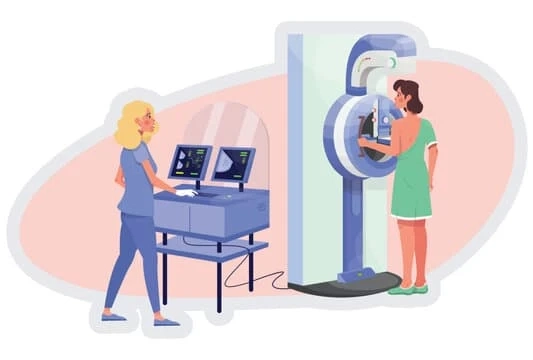
The mammography equipment looks like a box with a tube. The unit takes breast x-ray with some special accessories to limit the X-ray exposure to the breast only. The device has a team to hold and compress the breast. The radiologist can ask to position the breast so that he can capture the images from different angles.
Not all digital mammogram machines can perform Breast tomosynthesis imaging test.
Why Do Doctors Recommend A Mammogram?
- Mammography is performed either for screening or to make a proper evaluation. Women older than 40 can undergo mammograms if they have symptoms like breast skin thickening, nipple discharge, erosive sore of the nipple, or breast pain.
- Doctors recommend a mammogram to evaluate the breast tissues when the physical examination is inconclusive. Women with dense, lumpy, or very large breasts may be screened with mammography.
- Women with a high risk for breast cancer or a history of breast cancer should be screened.
There are some other reasons for mammography.
- If there is a new lump or thickening in the armpit
- If there is a change in the size or shape of your breast
- If there is a change in the breast, such as puckering, dimpling, a rash or redness of the skin
- Mammograms may be performed if there is fluid leaking from the nipple.
- If there is a change in the position of the nipple
- The mammography is an useful evaluation tool in the detection of abnormal growths that are confined to the milk ducts.
Mammography is conducted with other tests, such as a breast examination and an ultrasound scan in a diagnostic clinic. The patient may also have to go for a tissue biopsy for further evaluation.
Are Mammograms Painful?
A person undergoing a mammography test may feel uncomfortable due to the pressure on the breast tissue from the compression. It is even painful for some women. But the discomfort doesn\'t last long; if you feel intense pain, you can inform the radiologist immediately during the test.
The level of discomfort depends on a few factors, which include:
- The size and density of your breasts.
- How much do your breasts need to be compressed?
- Sometimes the breasts may be more tender & sensitive if you are having periods on the day of examination.
Why Is Breast Compression Required During a Mammogram?
Mammography exposes the breasts to small amounts of radiation, but the advantages outweigh the possible harm from the risk of radiation exposure. Radiologist compresses the breast during a mammogram test to:
- To evenly spread out the thickness of the breast so that all the tissues can be visualized properly.
- To capture the small abnormalities in the breast tissue which may be hidden by overlying tissue.
- The compression helps to allow a lower X-ray dose so that a thinner amount of breast tissue is captured.
- The person should hold the breast still so there is a minimum blurring of the image; a little motion can cause blurred images.
- The compression reduces the X-ray beam scatter to increase the sharpness of the picture.
- The radiologist can ask to change the positions to take the views from the top-to-bottom and an angle. The process is repeated for the other breast too.
- Compression is necessary for tomosynthesis imaging to minimize any kind of movement which degrades the picture.
A Final Say:
If you want a mammogram test, you can search for a mammography test price in Delhi for the nearest diagnostic centers. Delhi has some of the best mammogram screening diagnostic centers. Before the examination, you can find the breast mammography risks & precautions by visiting the website. You can call the health care executives who can guide you with all the related information regarding the mammogram. Nowadays, modern and state-in-art X-ray systems have minimized the effects of radiation by using controlled X-ray beams and other methods. Don\'t worry, and your body will receive minimal radiation exposure.


