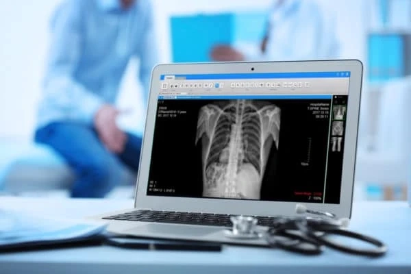
X-rays are a naturally occurring type of electromagnetic radiation that can generate images of internal tissues and structures in the human body. They are produced when charged particles collide with an obstacle. The internal structures in our body absorb X-rays based on their density.
For instance, the dense materials, like bone, appear white on the X-ray results. Since its inception in 1895, X-rays have changed how we look inside the human body. X-ray radiography is evolving quickly, thanks to digital technologies. Here are some common aspects to learn about X-ray radiography and the types of digital imaging.
What Is X-Ray Radiography?
X-ray radiography utilises ionising radiation in smaller doses to develop photographs of the human body’s internal structures. It is the oldest and most common type of medical imaging technology. From helping diagnose broken bones to identifying foreign substances in the body, X-rays are crucial to the healthcare sector’s efficiency.
The primary principle behind X-ray radiography lies in the differential absorption of X-rays by various tissues. Radiology technicians can capture a detailed illustration of your body’s internal structures. Some X-ray diagnosis tests may rely on a contrast material to produce better pictures of your bones and tissues.
The Advent Of The Digital X-Rays
The advent of digital X-rays is changing the entire radiography landscape in healthcare settings. It is ushering in a promising era of enhanced picture quality. Moreover, healthcare experts can access a wide range of flexible options to improve the clarity of pictures captured through digital X-machines. Some of the key advantages of digital X-ray radiography are as follows.
- Instant acquisition of pictures in high-resolution monitors
- Improves the quality of the photographs and their contrast
- Decreases exposure to radiation
- Digital X-rays can easily integrate with electronic medical records
The Types Of Digital Imaging
Now that you know about X-ray radiology, let’s discuss the types of digital imaging.
Fluoroscopy
Fluoroscopy is a real-time digital imaging technique that allows healthcare professionals to visualise the dynamic processes within the body. The procedure involves a continuous stream of X-rays passing through the patient.
Computed Radiography
Computer Radiography bridges the gap between conventional X-rays and digital X-rays. In this procedure, a phosphor plate stores the pictures captured by the X-rays. A laser beam scans this plate to convert analogue data into digital data.
Mammography
Mammography is a digital X-ray imaging technique that specifically detects breast abnormalities. It relies on low-energy X-rays to create detailed images of the breast tissue. Moreover, with mammography, doctors can recognise abnormal lesions.
Computed Tomography (CT)
In this procedure, the patient lies on the table and is placed in a ring-shaped scanner. The X-rays pass through the patient onto numerous detectors. The patient rotates slowly in the machine so the technicians can capture a series of slices to develop a 3D image. Note that the CT procedure involves the highest concentration of X-rays.
The world of X-ray radiography and digital imaging is undergoing an excellent transformation. In the future, numerous healthcare centers and medical facilities will integrate digital imaging techniques. The possibilities of X-ray radiography and digital imaging are boundless.


