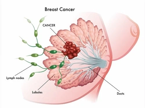
The breast cancer patient-derived xenografts model can not only faithfully retain the molecular phenotype and genotype changes of the patient\'s tumor but also reproduce the heterogeneity of the primary tumor, so it has been applied to the mechanism of tumor drug resistance and anti-tumor drug screening. This model maintains the biological characteristics of the patient\'s tumor gene expression pattern, mutation status, drug response, and tumor structure, and has an irreplaceable role in the translational medical research of clinical tumor treatment. PDX, as a new tool for the preclinical evaluation of tumor drug sensitivity, is attracting more and more attention. This article summarizes the types of experimental animal breast cancer models and the existing problems of PDX models.
Medicilon has established a variety of animal models in breast cancer disease, including the syngeneic model, xenograft model, PDX model, humanized tumor model, and orthotopic model. Now, Medicilon has the PDX models covering colon cancer, lung cancer, gastric cancer, breast cancer, liver cancer, and pancreas cancer. Our research on the PDX model includes molecular-level genotyping and pharmacological efficacy evaluation service of the orthotopic model, promising great predictions for clinical efficacy research.
What is a PDX mouse model?
In the PDX model, small tumors surgically excised from cancer patients are implanted into highly immunodeficient mice, allowing the tumors to grow and then transplanted into secondary recipient mice. Compared with the CDX model, the PDX model transplants intact tumor tissue into the recipient animal with the tumor structure and the relative proportion of cancer cells and stromal cells kept unchanged, resulting in enhanced consistency with human disease. [1] The patient-derived xenograft (PDX) model retains the cell morphology, tissue structure characteristics, and genetic characteristics similar to the patient to the greatest extent of the original tumor. The PDX model provides a new platform for studying the response of cancer patients to radiotherapy and chemotherapy, seeking new therapeutic targets, and improving prognosis, bringing personalized precision therapy research into a new stage.
Animal breast cancer model
Syngeneic model
The syngeneic model is to inoculate the cell line or tissue of breast cancer in experimental animals (such as rodents) into the same species or the same strain of immunologically normal animals. Some syngeneic models have been established, such as the mouse 4T1 model. The 4T1 cell line is isolated from a subpopulation system derived from the Balb/cf C3H mouse strain with spontaneous breast cancer. There is evidence that transplantation of the 4T1 mouse breast cancer cell line into immunocompetent mice of the same strain mimics the development and metastasis of human breast cancer compared to transplantation of human breast cancer cell lines (xenografts). It is more effective to observe the development and metastasis of tumors by injecting them into immune-deficient mice has been widely reported and utilized in the relevant literature, so the applicability of the same-strain mouse model with this cell line has been recognized and accepted by the academic community in this field.
Xenograft model
The xenograft model is to transplant human breast cancer cells or tissues into immunodeficient animals. Currently, three human breast cancer cell lines, MCF-7, T47-D, and MDA-MB-231, are most widely used in most preclinical studies of breast cancer. Most of the experimental animals are immunodeficiency strains of rodents, and the two most commonly used strains are severe combined immune deficiency (SCID) mice and nude mouse strains. The scientists studied the resistance of breast cancer to the continuous use of TM208 in human breast cancer xenograft mice. They identified the relationship between tumor pEGFR expression level and tumor growth inhibition. Some scientists have studied the implantation of benign and malignant tumor grafts into nude mice to create a nude mouse graft model to provide a preliminary screening tool for malignant tumor lesions through contrast agents. The specific method is to transplant malignant fibroma cells (cells and MDA-MB231 cells) and benign grafts with mitotic characteristics into both sides of nude mice simultaneously, and the success rate of transplantation can reach 90%. After five weeks, the sonogram showed that the volume of benign and malignant grafts reached 4 cm3 (about 2 cm in diameter), which made human tumors replicate well in nude mice. This research provides a basis for the treatment of human diseases. Xin et al. injected 200 μL MCF-7 cell suspension (5×10⁶) into the left thoracic mammary fat pad of female nude mice. The size of the tumor could reach 100-300 mm3 after a period of time, using this as an animal model for research of quercetin (Qu) in the treatment of breast cancer. [2]
Application of PDX model in breast cancer drug screening
PDX has enormous potential applications, including identifying new therapeutic targets and optimal treatment timing, analyzing and evaluating preclinical oncology drug combinations, biomarker identification, and resistance mechanisms. In some cases, preclinical research dominates clinical Experiments. For example, Schott et al. showed that in PDX models of chemotherapy-resistant metastatic breast cancer, drug inhibition of the Notch pathway reduced the number of tumor-initiating cells. For advanced breast cancer patients, preclinical research of the 0-secretase inhibitor MK-0752 combined with Docetaxel has been extended to phase 1b clinical trials. The key issue in drug development using PDX models is the degree of drug response, which reflects the clinical effectiveness of relevant patients. Many research groups have reported transplanted tumors and patient responses to drugs. There is consistency, although individualized treatment is challenging to evaluate clinical treatment, that is, the curative effect cannot be evaluated in a single way, but the basic standard for evaluation is that the tumor does not recur. In addition, breast cancer patients often receive multiple treatments (surgery, radiotherapy). Therefore, it is difficult to evaluate the efficacy of a single method. PDX models have been tried to detect individual responses to different drugs in patients with pancreatic cancer, and it has become a necessary detection step, which has guiding significance for individualized treatment.
However, even assuming stable drug sensitivity in the original tumor PDX model, this approach, first proposed 40 years ago, is challenging to apply to breast cancer. In fact, drug susceptibility testing is limited to individual patients, and the acquisition rate is low. Even after successful tumor transplantation, there is still a long interval for drug susceptibility results to guide clinical chemotherapy. But there are exceptions, such as triple-negative breast cancer cell subsets forming xenografts, which have a relatively high acquisition rate and a short delay time. Recently, a PDX model of a patient with metastatic triple-negative breast cancer could be formed within three weeks. Tumor, this patient has been guided by transplanted tumors for individualized treatment. [3]
Problems with PDX models
When applying the PDX model and analyzing the experimental results, we must clearly realize that the advantages of the PDX model are relative. Mice and humans are always two different species, and the differences between species cannot be completely eliminated. Since the microenvironment of human tumor tissue transplantation in the PDX model is still mouse-derived but not human-derived, some researchers added human-derived immune tissues or cells to immunodeficient mice to establish a new type of "human-in-mouse" model. Zheng Mingjie et al. transplanted human mammary gland tissue into the abdomen of SCID mice subcutaneously and injected human breast cancer cells into the surviving transplanted breast tissue when the transplanted tissue grew to 80% confluence one week after the operation. The results show that human breast cancer cells can grow better in the human breast microenvironment. This new human breast mouse model better simulates the interaction between the human microenvironment and tumor cells. People try to introduce human factors into the PDX model; the purpose is to make the test model better reflect the actual situation of the patient\'s tumor, and the clinical test results obtained by relying on this type of model can be better transformed into clinical effective treatments.
At the same time, in order to reduce the rejection reaction and improve the survival rate of transplantation by transplanting human-derived tissues into mice, immunodeficiency mice are mainly used to establish models, which leads to the lack of an immune microenvironment in-vivo. In addition, the nude mice commonly used to prepare PDX models only lack T cells, and the B cells and NK cells in the body also play a specific screening role so that some heterogeneity of the primary tumor may be lost in the in vivo passage. It is essential to be aware of the absence of specific phenotypes when analyzing PDX model results. Mice with a higher degree of immunodeficiency should be selected, such as NOD-SCID mice, which are currently widely used, non-obese diabetic/severe combined immunodeficiency mice, which have T, B, NK cell and complement deficiencies, and more suitable for transplantation of human tumor specimens. Similarly, NSG mice are also triple immunodeficiency of T, B, and NK cells. At the same time, their macrophage function and complement hemolytic activity are all reduced, which is more conducive to reducing the screening effect of the body\'s immune system on foreign grafts and avoiding the risk of primary tumors. Deletion of specific phenotypes. Studies have found that the IL2rg gene is closely related to the body’s immune function. After the gene is knocked out, the immune function of the mouse body is severely reduced, especially the activity of NK cells is almost lost. Someone backcrossed IL2rg-/- mice with NOD-SCID mice, and Immunodeficient mice with both advantages were obtained. Generally, immunodeficient mice have a shorter survival time due to the high degree of deficiency, which affects the growth of transplanted tumors. The newly reported B-NSG mice, combined with the background characteristics of NOD-SCID-IL2rg, not only have a severe immunodeficiency phenotype, but also live longer than NOD-SCID mice, with an average of 1.5 years. It can be seen that the development of PDX models is closely related to the research progress of immunodeficient animals. Since different mouse models differ in the degree of immunodeficiency, survival time, operation requirements, and living environment, we should choose the corresponding mouse model according to the actual needs.
In addition, the cost of PDX model modeling is high, the cycle is long, and only the primary transplantation takes more than 2 to 3 months. The technical requirements of the operation are high, and the specimens need to cooperate closely with surgeons, histologists, and researchers after they are obtained from the operating room. At the same time, due to the limited number of patient-derived tumor tissues and the restrictions on their use by medical ethics, the number of existing primary models is small, and the passaged samples need to be tested by histopathology to determine their consistency with the original samples. After undergoing this series of precise and complex operations, the average tumor formation rate of PDX models is only about 25%. [4]
Medicilon\'s PDX Model
Now, Medicilon has the PDX models covering colon cancer, lung cancer, gastric cancer, breast cancer, liver cancer, and pancreas cancer. Our research on the PDX model includes molecular-level genotyping and pharmacological efficacy evaluation service of the orthotopic model, promising great predictions for clinical efficacy research.
[1]. Jialiang Hu,et al. Pharmaceutical Biotechnology. PDX Model-Aided Cancer Disgnosis and Treatment. 2022,29(05),528-532 DOl:10.19526/j.cnki.1005-8915.20220517
[2]. Rifi Li et al. Research progress in experimental animal models of breast cancer. Chinese Journal of Comparative Medicine. 2018,28(02):113-118.
[3]. Lijun Ma et al. Recent Progress in Patient-Derived Tumor Xenograft Model of Breast Cancer. Scientia Sinica(Vitae). 2016,46(06):695-704.
[4]. Binquan Hu et al. The Advantages and Disadvantages of Human PDX Transplantation Model. Laboratory Animal Science. 2015,32(05):59-62.


